Genetic Characterization of Rat Hepatic Stellate Cell Line HSC-T6 for In Vitro Cell Line Authentication

https://doi.org/10.3390/cells11111783
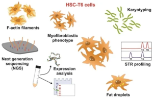
Abstract Immortalized hepatic stellate cells (HSCs) established from mouse, rat, and humans are valuable in vitro models for the biomedical investigation of liver biology. These cell lines are homogenous, thereby providing consistent and reproducible results. They grow more robustly than primary HSCs and provide an unlimited supply of proteins or nucleic acids for biochemical studies. Moreover, they can overcome ethical concerns associated with the use of animal and human tissue and allow for fostering of the 3R principle of replacement, reduction, and refinement proposed in 1959 by William M. S. Russell and Rex L. Burch. Nevertheless, working with continuous cell lines also has some disadvantages. In particular, there are ample examples in which genetic drift and cell misidentification has led to invalid data. Therefore, many journals and granting agencies now recommend proper cell line authentication. We herein describe the genetic characterization of the rat HSC line HSC-T6, which was introduced as a new in vitro model for the study of retinoid metabolism. The consensus chromosome markers, outlined primarily through multicolor spectral karyotyping (SKY), demonstrate that apart from the large derivative chromosome 1 (RNO1), at least two additional chromosomes (RNO4 and RNO7) are found to be in three copies in all metaphases. Additionally, we have defined a short tandem repeat (STR) profile for HSC-T6, including 31 species-specific markers. The typical features of these cells have been further determined by electron microscopy, Western blotting, and Rhodamine-Phalloidin staining. Finally, we have analyzed the transcriptome of HSC-T6 cells by mRNA sequencing (mRNA-Seq) using next generation sequencing (NGS).
Original Publication (Open Access)
Genetic Characterization of Rat Hepatic Stellate Cell Line HSC-T6 for In Vitro Cell Line Authentication.
I Nanda, C Steinlein, T Haaf, EM Buhl, DG Grimm, SL Friedman, SK Murer, SK Schröder, R Weiskirchen
Cells, 2022 (https://doi.org/10.3390/cells11111783)
